Back Of Neck Anatomy - Torso Model Unisex Opened Neck And Back 3b Scientific 1000193 B19 / This muscle is controlled by the third and fourth cervical.
byAdmin•
0
Back Of Neck Anatomy - Torso Model Unisex Opened Neck And Back 3b Scientific 1000193 B19 / This muscle is controlled by the third and fourth cervical.. Working in pairs on the left and right sides of the body, these muscles. The motion of the muscles of the neck are divided into four. The back anatomy includes some of the most massive and functionally important muscles in the human body. Arteries of the neck model 9 photos of the arteries of the neck model arteries in the back of the neck, arteries in the neck and head, arteries veins neck, blocked arteries neck, clogged arteries neck, main arteries neck, major arteries neck, neck, arteries in the back of the neck, arteries in the neck and … anatomy face and neck side view Still, many individuals pay far too little attention to them.
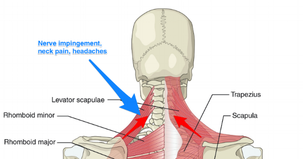
Rarely, neck pain can be a symptom of a more serious problem. The image below to shows all the major back muscles (as well as some neck muscles): The back of the neck is mostly comprised of muscles, as well as the spine. In particular, the levator scapulae muscle is susceptible to injury. They move the head in every direction, pulling the skull and jaw towards the shoulders, spine, and scapula.
Pin On Fitness Back Workouts from i.pinimg.com The suboccipital muscles act to rotate the head and extend the neck.rectus capitis posterior major and rectus capitis posterior minor attach the inferior nuchal line of the occiput to the c2 and c1 vertebrae respectively.obliquus capitis superior also extends from the occiput to c1 while obliquus capitis inferior originates from c2 and. Seek medical care if your neck pain is accompanied by numbness or loss of strength in your arms or hands or. They ultimately drain into the deep lymph nodes. They move the head in every direction, pulling the skull and jaw towards the shoulders, spine, and scapula. Neck muscles can be strained from poor posture — whether it's leaning over your computer or hunching over your workbench. The back of the neck is mostly comprised of muscles, as well as the spine. See anatomy of the head and neck stock video clips. The trapezius, commonly referred to as the traps, are responsible for pulling your shoulders up, as in shrugging, and pulling your shoulders back during scapular retraction.
Osteoarthritis also is a common cause of neck pain.
The neck triangles are actually spaces bordered by the neck muscles. The muscles of the neck run from the base of the skull to the upper back and work together to bend the head and assist in breathing. They move the head in every direction, pulling the skull and jaw towards the shoulders, spine, and scapula. See anatomy of the head and neck stock video clips. It runs from the neck to the upper back. Pain and dysfunction from injuries or conditions that impact the joints, muscles, and other structures can easily spread from the neck to the shoulder(s) and from the shoulder(s) to the neck. The neck and shoulders are complex and interconnected areas, and medical problems that affect one often affect the other, as well. The spine runs from the base of your skull down the length of your back, going all the way down to your pelvis. Muscle head anatomy vocal organ diagram female neck anatomy neck wireframe head neck human anatomy head artery anatomy face pharynx vector neck degree head anatomy 3d. Back of neck anatomy : These two ligaments connect and support the spine from the neck to the lower. Cervical spine anatomy video the cervical spine has 7 stacked bones called vertebrae, labeled c1 through c7. The superficial lymph nodes of the head and neck receive lymph from the scalp, face and neck.
The back anatomy includes some of the most massive and functionally important muscles in the human body. The anterior, and the posterior, triangles of the neck. It runs from the neck to the upper back. The skull is a strong, bony capsule that rests on the neck and encloses the brain. It is made up of bones, discs, muscles, ligaments, nerves and tendons.
Preventitive Care Blog Back Pain And Headache Specialist Burke Va Nova Headache Chiropractic Center from images.squarespace-cdn.com The neck is connected to the upper back through a series of seven vertebral segments. In addition, in this region we also find the major cranial and spinal nerves that connect the central nervous system to the organs, skin, and muscles of the head and neck. In this section, learn more about the anatomy of the muscles of the neck. The top of the cervical spine connects to the skull, and the bottom connects to the upper back at about shoulder level. Extending from underneath the chin, to the posterior aspect of the head. In particular, the levator scapulae muscle is susceptible to injury. Neck anatomy nerves picture there are 8 spinal nerves that originate from the cervical spine. Below the neck, holding the tooth into the bone, is the root of the tooth.
In addition, in this region we also find the major cranial and spinal nerves that connect the central nervous system to the organs, skin, and muscles of the head and neck.
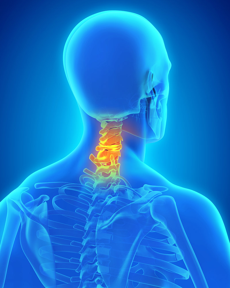
Still, many individuals pay far too little attention to them. Anatomy of respiratory system 12 photos of the anatomy of respiratory system anatomy and histology of the respiratory system ppt, anatomy and physiology of respiratory system with pneumonia, anatomy and physiology respiratory system worksheets, detailed anatomy of respiratory system, human anatomy respiratory. Cervical spine anatomy video the cervical spine has 7 stacked bones called vertebrae, labeled c1 through c7. Causes of neck pain and how to manage the pain in basic terms, the neck (cervical spine) joins the shoulders and chest to the head. The anterior triangle of the neck is made by the anterior border of the sternocleidomastoid muscle, the inferior border of the mandible and the midline of the neck. The neck triangles are actually spaces bordered by the neck muscles. They start at the top of the neck and go down to the tailbone. The larynx is located where the pharynx, the back of the mouth and nasal cavity, divides into the trachea (the tube that carries air to the lungs) and the esophagus (the tube that carries food to. Neck anatomy explained the neck begins at the base of the skull and connects to the thoracic spine (the upper back). They are arranged in a ring shape; Pain and dysfunction from injuries or conditions that impact the joints, muscles, and other structures can easily spread from the neck to the shoulder(s) and from the shoulder(s) to the neck. These two ligaments connect and support the spine from the neck to the lower. Neck anatomy nerves picture there are 8 spinal nerves that originate from the cervical spine.
Located at the back and side of the neck, the levator scapulae muscle connects the neck's cervical spine with the shoulder. The nerves of the head and neck include the most vital and important organs of the nervous system — the brain and spinal cord — as well as the organs of the special senses. The rotation function takes the head into the opposite side to which this neck and shoulder muscle is located. It is composed of three parts: Working in pairs on the left and right sides of the body, these muscles.
A Literal Pain In The Neck The Center from www.thecenteroregon.com The neck and shoulders are complex and interconnected areas, and medical problems that affect one often affect the other, as well. Neck anatomy explained the neck begins at the base of the skull and connects to the thoracic spine (the upper back). The larynx is located where the pharynx, the back of the mouth and nasal cavity, divides into the trachea (the tube that carries air to the lungs) and the esophagus (the tube that carries food to. The anterior triangle of the neck is made by the anterior border of the sternocleidomastoid muscle, the inferior border of the mandible and the midline of the neck. Extending from underneath the chin, to the posterior aspect of the head. The anterior, and the posterior, triangles of the neck. Cervical spine anatomy video the cervical spine has 7 stacked bones called vertebrae, labeled c1 through c7. They ultimately drain into the deep lymph nodes.
The neck muscles, including the sternocleidomastoid and the trapezius, are responsible for the gross motor movement in the muscular system of the head and neck.
The neck is one of the most complex and intricate structures in our body and includes the spinal cord, which sends messages from the brain to the rest of the body. The neurocranium (cranial vault) and the viscerocranium (facial skeleton). Back of neck anatomy : These two ligaments connect and support the spine from the neck to the lower. Anatomy of respiratory system 12 photos of the anatomy of respiratory system anatomy and histology of the respiratory system ppt, anatomy and physiology of respiratory system with pneumonia, anatomy and physiology respiratory system worksheets, detailed anatomy of respiratory system, human anatomy respiratory. Below the neck, holding the tooth into the bone, is the root of the tooth. Neck anatomy explained the neck begins at the base of the skull and connects to the thoracic spine (the upper back). The top of the cervical spine connects to the skull, and the bottom connects to the upper back at about shoulder level. Related posts of anatomy of the back of the neck anatomy of respiratory system. Muscle head anatomy vocal organ diagram female neck anatomy neck wireframe head neck human anatomy head artery anatomy face pharynx vector neck degree head anatomy 3d. Extending from underneath the chin, to the posterior aspect of the head. This muscle is controlled by the third and fourth cervical. Causes of neck pain and how to manage the pain in basic terms, the neck (cervical spine) joins the shoulders and chest to the head.